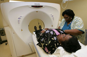
 (Justin Sullivan/Getty Images)
ROCKVILLE, MD Just before Christmas, 41-year-old Michael Kelley decided he wanted a whole-body imaging exam, the heavily advertised service touted on television by celebrities like Oprah Winfrey. He didn’t smoke, wasn’t overweight, and didn’t have elevated cholesterol. “I’m pretty normal for a guy my age,” he said.
No matter. The electrical engineer scheduled a full-body X-ray computed tomography or CT scan at Virtual Physical, a radiology clinic located in a glass-enclosed office building on a busy commercial strip not far from the headquarters of the National Institutes of Health. The clinic’s name, plastered in large red letters on the building’s exterior, served as a billboard aimed at cars exiting the high-end shopping mall across the street.
About an hour after checking in, Kelley left the clinic clutching a manila envelope with high-resolution 3-dimensional images of most of his major body systems, including the insides of the major coronary arteries pumping blood to and from his heart. “They said I was fine, no plaque,” he said. Kelley paid $1,400 for a CT scan to confirm what he and his doctor already knew – he was perfectly healthy.
High-tech medical imaging advances, along with robotics, new devices, body parts, and pharmaceuticals have changed almost every aspect of health care and improved the quality of life for young and old alike.
The U.S. leads the world in creating state-of-the-art diagnostic and therapeutic treatments with the potential to work miracles in millions of patients. But the miracles come at a stiff price. Unlike other industries where technology has helped lower costs by eliminating waste and increasing productivity, technology has become a major factor in increasing health care costs. In a January 2008 report, the Congressional Budget Office concluded that “roughly half of the increase in health care spending during the past several decades was associated with the expanded capabilities of medicine brought about by technological advances.”
One reason for this dichotomy is the overuse and misuse of technology as preventive medicine.
The U.S. Preventive Services Task Force recommends against routine imaging screening for heart disease in healthy people like Kelley because many of those scans show false positives, sending people for further tests and procedures that needlessly drive up health care costs. CT scans also heighten one’s cancer risk because of high radiation levels.
Even so, the tests have proliferated in the half decade since advanced CT scanning machines hit the market. Since 2001, the number of scanning machines in the U.S. has increased by over 20 percent, while the number of scans taken each year has gone from one for every seven persons to almost one for every four. Yet there’s no evidence that the additional invasive procedures that result from this large increase in screening are preventing serious heart disease.
The cardiologists and radiologists who rely on these scanners claim the spectacular images produced by the newest machines eliminate many of the unnecessary invasive procedures given to people with symptoms of heart disease. Yet there’s no evidence that the high-tech images have reduced the number of patients needing exploratory catheterization, a delicate and risky procedure. In fact, the rate of invasive coronary artery procedures has actually gone up in the past decade
Scanning for Business
In 2008, the Sutter Tracy Community Hospital in the northern part of California’s Central Valley bought the pitch. It purchased General Electric’s latest $1.4 million “LightSpeed” CT scanning machine, which records 64 high-resolution images or “slices.” According to the website RadiologyInfo, “CT imaging is sometimes compared to looking into a loaf of bread by cutting the loaf into thin slices. When the image slices are reassembled by computer software, the result is a very detailed multidimensional view of the body’s interior.”
“This CT scanner is five times faster and produces higher resolution images than the previous CT scanner we installed five years ago,” David Bowlsby, the hospital’s imaging manager, said in a press release. “It also has the ability to capture pictures of a beating heart, something our last CT scanner couldn’t do.”
Tracy Sutter’s purchase came at the tail end of a huge boom in sophisticated imaging equipment sales, which has slowed in the past year and a half because of the recession and public concern about rising health care costs. In the four years after the 2003 introduction of the 64-slice CT scanning machine, the number of CT scanners in the U.S. jumped by nearly 2,000, a 23 percent increase. “It was double-digit growth until the past two years,” said Morningstar analyst Daniel Holland. “Sales have been down of late because the latest and greatest scanners cost well over $1 million and people sense we’re being overdiagnosed.”
Manufacturers like GE, Toshiba, Philips and Siemens market these expensive machines to large hospitals and independent clinics, the latter often owned by cardiologists and radiologists who cater to the worried well. When deployed in a fee-for-service marketplace with few brakes on spending, there are powerful economic incentives to use them as widely as possible, especially when there’s a very high initial or fixed cost for their creation and installation. These high capital costs compel some providers to use the technologies on patients for whom less costly drugs, tests and procedures would suffice.
“What is driving costs in health care is the development and adoption of new technologies that offer tremendous hope in some patients, but also present the tremendous opportunity to be overused,” said Amitabh Chandra, a professor of public policy at the Kennedy School of Government at Harvard University. “Once I have the facility, I have a powerful incentive to do as many as possible.”
Moore’s Law Redux
Like computer processing speed and digital camera megapixels, CT scanning capability, measured by the number of computed image “slices,” seems to double every few years. What started with black and white one-slice machines in the 1970s has developed into software-driven imaging machines that can deliver color images like those seen in a 3-D movie. But unlike computers and cameras, the better the CT scan image, the greater the cost. Are these ever-improving images worth the price?
Julie Miller, an interventional cardiologist at Johns Hopkins University Medical Center, thinks they are. She has performed thousands of coronary procedures in the past decade. But only recently has she been able to view lifelike images of the insides of her patients’ arteries before scheduling their operations.
In 2007 the elite hospital installed one of the most advanced medical imaging devices in the world, a 320-slice computed tomographic (CT) X-ray scanning machine. One of only 200 in the world, the Toshiba Aquilion One tipped the price scale at $2.2 million, well above the cost of the 64-slice machine it hopes to replace.
Sitting in front of a work station in the cardiology department’s computer lab, she clicked on several patient records to show what a fivefold increase in scanning power could reveal. The new machine provided far sharper images of the major coronary arteries feeding the heart more like a Disney animation than the flat images of older machines. The new machine also reduced scanning time to a few seconds, allowing more patients to take the test, and exposing them to less radiation.
At about $400 to $1,000 per scan, the advanced technology behind 64-slice CT angiography held out the promise of achieving one of the holy grails of cardiology a non-invasive test that will help doctors determine if people at high risk or already showing symptoms of severe heart disease (like persistent chest pains) need to undergo angioplasty and stenting for coronary blockages, which can cost $50,000 or more. Older tests result in unnecessary cath lab referrals in about 30 percent of cases.
“This is almost a replacement technology for nuclear and stress echocardiograms and even for standard stress testing,” she said. “We’re going to start seeing catheterization only for when you really need to do revascularization.”
“It’s an excellent test for excluding the presence of coronary artery disease,” said Geoffrey Rubin, professor of radiology at Stanford University and chair of the cardiovascular imaging committee for the American College of Radiology. “Given that there are a lot of negative angiograms performed in this country every day, the use of CT angiography to prevent those far more expensive and far more risky interventions is telling.”
With strong backing from both specialist physician groups, the U.S. has installed nearly twice as many CT machines per capita than the average industrialized nation: 34 per million people in 2007, compared to 15 per million elsewhere, according to the Organization for Economic Cooperation and Development. Cardiologists’ revenue from Medicare spending on imaging services rose to 36 percent of total revenue in 2006, up from 23 percent in 2000, according to a 2008 Government Accountability Office report.
To date there is no evidence that CT scans have reduced heart attacks or cardiovascular disease deaths, or even reduced catheterization lab use. In 2006 there were 1.6 million invasive procedures (known as percutaneous coronary interventions or PCIs, which encompass both the exploratory catheterization angiograms and the angioplasty and stenting to prop open the damaged arteries that follow in the catheter’s wake), compared to 1.4 million in 1995. The rates edged up, too, rising to 53.9 per 10,000 persons in 2006 from 53.2 per 10,000 in the middle of the last decade.
“The pictures look beautiful, but there’s still no outcome data to speak of,” said Rita Redberg, a University of California at San Francisco cardiologist and editor of the Archives of Internal Medicine. She believes the proliferation of machines, coupled with Medicare reimbursement, has led cardiologists and radiologists to give heart scans to people like Mike Kelley, with few or no serious cardiovascular risk symptoms. Yet some of those scans reveal calcification or plaques that, upon catheterization, turn out to be insignificant. These false positives, Redberg says, undermine the usefulness of the screening test.
Positively False
Early in 2008, a 52-year-old nurse at a suburban Cleveland hospital began complaining of persistent chest pains. Her physician ordered a CT scan, which turned up calcification in one of the major arteries feeding her heart. She was immediately sent to the catheterization lab to see if there was a severe blockage behind the calcification, which there often is. In this case, there wasn’t. The CT scan had turned up a false positive.
As the interventional cardiologist carefully snaked the catheter through the artery, he accidentally perforated the nurse’s left main artery, leading to emergency open heart surgery. They had to crack open her chest to repair the damage, followed by intensive recovery in the ICU. The nurse recovered, but not before her heart was permanently damaged from the temporary reduction in blood flow. In the following year, she suffered at least two heart attacks that lead to hospitalization and was eventually recommended for a heart transplant. “It was all precipitated by a study she didn’t need,” said Matthew Becker, the attending cardiologist at the Cleveland Clinic Foundation who treated one of her subsequent heart attacks. Artery perforation is a rare event during catheterization procedures, but does occur in anywhere from 1 to 3 in 1,000 cases.
A study that appeared in December 2008 in the Journal of the American College of Cardiology showed that over 50 percent of all CT detected coronary obstructions were false positives. “This high false-positive rate has potentially serious implications, leading to unnecessary and potentially risky procedures that threaten to accelerate already excessive health care costs,” said an accompanying editorial by Steven Nissen, chair of the Cleveland Clinic’s cardiovascular medicine division, which conducts research for numerous drug and device manufacturing companies.
Despite those risks, when Medicare announced a plan in December 2007 to rein in spending on CT angiography by requiring clinical trials and limiting CT scan use to patients with symptoms of heart disease, it was bombarded with 649 protest letters from cardiologists and radiologists, their professional societies, patient advocacy groups and equipment manufacturers. Even Congress got involved, with 79 members of the House sending a letter noting their opposition to Kerry Weems, the acting administrator of the Center for Medicare and Medicaid Services. The agency backed off.
A Smoker’s Gun?
Cardiologists are not alone in misusing high-tech medical technologies. In October 2006, stunning news for smokers was splashed across the front pages of many leading newspapers. A report in the New England Journal of Medicine showed annual CT chest scans increased 10-year survival for those identified with early-stage lung cancer to 88 percent, up from 5 percent previously. The study, partially funded by the tobacco industry, intimated annual screening of asymptomatic smokers would achieve near miraculous results.
“In a population at risk for lung cancer, such screening could prevent some 80% of deaths from lung cancer,” the study’s authors wrote in the prestigious medical journal.
Other researchers immediately attacked the optimistic finding. A team of Dartmouth University scholars pointed out that an early diagnosis of lung cancer does not necessarily mean treatment would be any more successful, or that survival rates would improve. Steve Woloshin, one of the Dartmouth authors, says that it’s possible that these patients would not live even a day longer. Many feared a repeat of the experience with chest X-rays for smokers, which in clinical trials succeeded in identifying many small tumors that were surgically removed. Later it was discovered that the surgery did not affect their long-term survival. They often found cancerous growths that were never destined to become life-threatening.
“They found cancers earlier, [but] it did not change mortality from lung cancer and it did not reduce the severity of the cancers,” said William C. Black, a radiologist at the Dartmouth Hitchcock Medical Center. What it did find, according to Black, was the X-rays picked up insignificant, benign cancers. The X-rays also found small but aggressive cancers that remained impervious to treatment despite their early detection.
The National Institutes of Health has funded a clinical trial to provide a definitive answer to whether CT screening of smokers will perform better than traditional X-rays in reducing lung cancer mortality. It has been underway since 2002. The National Lung Screening Trial enrolled over 50,000 patients at more than 30 sites across the U.S. While the results of the study, which concluded last year, won’t be known until the end of 2011, it’s likely the benefit of CT screening for smokers will be minimal at best.
“You can assume there’s no huge trend in the data because the data safety monitoring board didn’t shut the study down,” said Black, whose center is participating. If the study showed a real advantage to CT scans over chest X-rays, the NIH would have given them to everyone.
Meanwhile, a preliminary finding from a government-funded study, released in mid-2009 at the American Society of Clinical Oncology’s annual meeting, showed the significant risk of giving routine CT scans to smokers. Once again, the scans recorded false positive findings of cancer-like growths in one out of every three patients screened, twice the rate of false positives turned up on older chest X-ray technology.
While no insurers, including Medicare, currently pay for CT scanning for the nation’s 46 million smokers, they do pay for the biopsies and excisions of the small lesions that are turned up by the exams. That’s why there’s no shortage of cancer centers across the country starting to push CT scanning for middle-aged smokers, perhaps as a loss leader for their other cancer treatment services. The International Early Lung Cancer Action Program, which backs routine CT scanning for smokers, lists 42 centers in 16 states offering the screening tests.
The Mother of all Machines
Medical imaging machines range from relatively simple X-ray and ultrasound to more sophisticated CT scans and nuclear positron emission tomography, or PET scans. PET is an investment of around $2 million and is part of the growing field of nuclear medicine. But the cost of the top-of-the-line PET machine pales by comparison to the $140 million proton beam therapy machine, which promises to deliver cancer-killing radiation to cancerous tumor cells without harming surrounding tissue. For men diagnosed with prostate cancer, this machine could be their miracle.
The popularization of prostate specific antigen (PSA) testing in the 1990s led to a dramatic increase in prostate cancer diagnoses. In 2009, more than 192,000 cases were estimated by the American Cancer Society. PSA tests have triggered an upsurge in every form of treatment: surgery, radiation and drug therapy.
Despite all the early diagnosis and treatment, the mortality rate remains stubbornly high, with over 27,000 deaths annually, down only modestly from its previous peak. Two studies released earlier this year help explain why. A U.S. study showed no reduction in the prostate cancer death rate after mass PSA screening, while a European study showed modestly lower mortality in those screened.
A significant finding in both studies, on the other hand, showed extensive overdiagnosis and overtreatment, according to Steven Woolf, a preventive health specialist at Virginia Commonwealth University.
Given the debilitating side effects of all the current forms of treatment, which include impotence and incontinence, it’s easy to understand why some men choose “watchful waiting” over aggressive treatment of the disease. But they’re still in the minority. The large majority opt for medical or surgical treatment and suffer the long-term consequences.
That’s created a major opportunity for providers whose treatments claim to avoid major side effects, which in a few communities now includes the most expensive medical device ever created the proton beam therapy machine. There are currently seven operating in the U.S. with four more under construction.
The machine requires building a cyclotron in a football field-sized facility that can deliver high-energy particles to a medical device. While originally touted for tumors of the head and neck that threaten sensitive nerves, its largest use to date has been among prostate cancer patients who fear the side effects of other interventions, including conventional radiation. The effect of proton beam radiation on cancer is no different than other forms of radiation, according to Hadley Ford, the chief executive officer of ProCure, a four-and-a-half-year-old startup company that has opened one center in Oklahoma and has another under construction in suburban Chicago. “The difference is how much damage I do,” Ford says.
He might be right, or he might be hyping a very expensive therapy that promises more than it delivers. A recent Agency for Healthcare Research and Quality review of clinical studies failed to turn up any evidence that supported his claim of fewer side effects. Additionally, “No study found that charged particle radiotherapy is significantly better than alternative treatments with respect to patient-relevant clinical outcomes,” the AHRQ review said.
Proof Positive
It may be a long time before high-cost proton beam therapy is tested against existing treatments. Ford said it would be unethical for his firm to conduct one. “You’d have to sit down with a patient and explain that in one [arm of the trial] you’d get more radiation,” he said. “What am I going to do, force him to go get more radiation?”
“There is no ethical roadblock,” countered Howard Brody, director of the Institute for the Medical Humanities at the University of Texas in Galveston. “To say you can’t begin a clinical trial because you’d be denying people a safer alternative is to assume you know what you have yet to prove.”
That same argument has been used before, pointed out Alan Garber, a health care economist at Stanford University and a former chairman of an advisory group on Medicare payment policy. High-dose chemotherapy, bone marrow transplants for breast cancer and lung volume reduction surgery for emphysema were used for years without supportive clinical trials because proponents presumed the interventions worked and they considered such trials unethical. However, government-funded investigators had no problem getting approval to conduct trials from institutional review boards, which are charged with reviewing trial methods and protocols to ensure they won’t harm patient safety. Those clinical trials eventually proved the three technologies “weren’t any better and they almost all died out,” Garber said. “Proton beams? We don’t know whether it works any better than any other technology. We [only] have a rationale that it is better.”
In the long run, technology may in fact reduce costs. When robotic doctors are able to perform micro-surgeries; when arm and leg replacements function as well if not better than the original parts; when pharmacology replaces more expensive treatments and therapies, the U.S. may actually be able to use technology to bend the cost curve of health care downward. Until that time comes, we’re stuck with ever-increasing costs and left to wonder whether the investment is greater than the payoff.
“Our biggest problem comes when a technology has real benefit for some people and may even save some lives,” said Jon Skinner, a professor of health economics at Dartmouth University. “[Then] it spreads out to more and more people where there’s not a lot of benefit. By the time the studies come out, it’s already become usual care.
“Once physicians are invested in a particular approach,” he said, “it’s hard to get them to change.”


